Blood is considered a connective tissue for two basic reasons: (1) embryologically, it has the same origin (mesodermal) as do the other connective tissue types and (2) blood connects the body systems together bringing the needed oxygen, nutrients, hormones and other signaling molecules, and removing the wastes. In circulating blood two different cell types are found: enucleated erythrocytes or red blood cells and nucleated leukocytes or white blood cells. We will study their histology in blood smears.
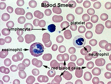
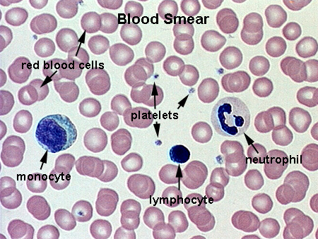
RBCs are small (7 um) cells lacking a nucleus. They stain red with eosin and due to their concave shape have a lighter staining center. Numerous examples are found in the images below.
The WBCs are divided into two groups: (1) granular leukocytes, containing distinctive cytoplasmic granules, including neutrophils, eosinophils and basophils and (2) agranular leukocytes, without granules, including monocytes and lymphocytes.
Neutrophils (10-12 um diameter), also called polymorphonuclear leukocytes (PMNs) or polymorphs, have nuclei with 2-5 lobes connected by thin chromatin threads. Their cytoplasm contains 2 types of granules; small neutrophilic granules (which give the cell a lavender hue and purplish-red azurophilic granules which are lysosomes (Neutrophil).
Eosinophils are larger than PMNs (12-17 um diameter), have a bilobed nucleus, and large eosinophilic (red-orange) granules in their cytoplasm (Eosinophil).
Basophils, which represent only 0.5% of the WBCs are difficult to find. They are 10-12 um diameter, with a large, irrgeularly shaped nucleus (the least condensed chromatin of all granulocytes). The cytoplasm is dominated with large dark blue (basophilic) granules (Basophil).
Monocytes are the largest WBC (14-20 um diameter) with variable nuclear morphology from ovoid to horseshoe shaped. The cytoplasm stains a light blue and contains a few pale azurophilic (lysosomal) granules (Monocyte).
Lymphocytes are a population consisting of both B- and T-lymphocytes. In blood smears they have the same morphology. One can see both small (slightly larger than an Rbc 7-8 um) and medium to large (up to 12 um); these represent inactive and activated lymphocytes respectively. Histologically, lymphocytes have very dense nuclei which occupies most of the cell with a thin rim of bright basophilic cytoplasm (Lymphocytes).
Platelets are fragments of cytoplasm detached from Megakaryocytes in the bone marrow. They aggregate together during the process of blood coagulation and clotting. Platelets are small (2-5 um), have no nucleus and are ovoid shaped. They have a dark staining core (granulomere) with a clear outer region (hyalomere) (Platelets).
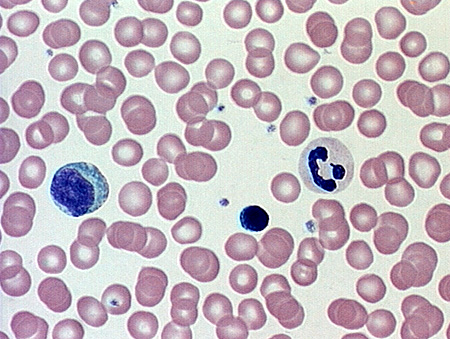
See if you can identify 5 different blood cells in each of the following fields:
Blood Smear 1 Answer
Blood Smear 2 Answer
Hemopoiesis is the continual production of new blood cells. There are two kinds of hemopoietic tissue: (1) myeloid tissue or bone marrow where RBCs, granular WBCs, platelets, monocytes are produced and (2) lymphatic tissue - thymus, spleen, lymph nodes, where lymphocytes are made.
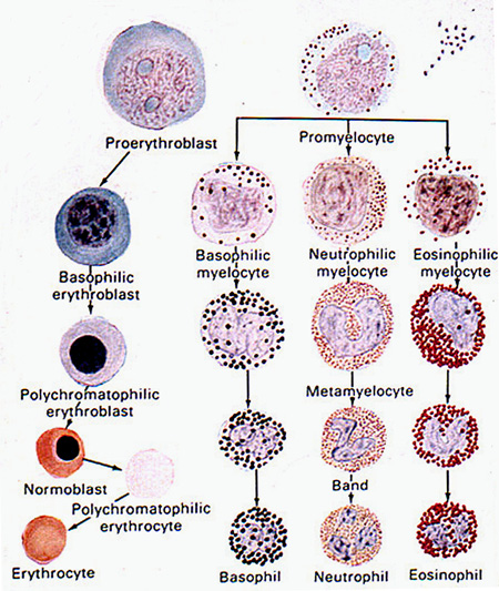
All blood cells have a common pluripotent stem cell which develops into each type in clonal units. We will focus on the morphological recognizable stages. During erythropoiesis, the large stem cells first accumulated ribosomes and then decrease in size, condensing their nuclei and synthesizing hemoglobin. A summary of the developmental stages are found in this diagram (Hemopoiesis)
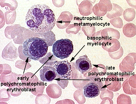
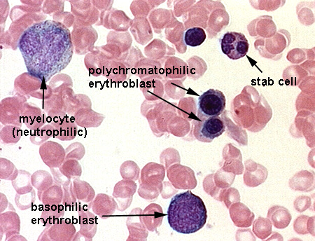
Basophilic erythroblasts are large cells (12-15 um) with a large nucleus beginning to condense. The cytoplasm, full of free ribosomes, stains intensely basophilic (Erythropoiesis 1).
Polychromatophilic erythroblasts are formed as the nucleus becomes more condensed (a soccer ball-like morphology) and hemoglobin begins to accumulate in the cytoplasm. The combined staining of the basophilic ribosomes and acidophilic hemoglobin give these cells a grayish cytoplasm (Erythropoiesis 2 and Erythropoiesis 3).
Normoblasts are approximately the size of mature RBCs and stain almost the same since most ribosomes have been lost as more hemoglobin accumulates. The retain a small highly condensed (pycnotic) nucleus (Erythropoiesis 4).
Polychromatophilic erythrocytes or reticulocytes are formed when a normoblast loses its nucleus. However, these cells still retain some ribosomes and combined with the hemoglobin, some polychromatic staining (pink-blue cytoplasm) is observed (Erythropoiesis 3).
This process occurs in bone marrow along side erythropoiesis with each type (eosinophil, basophil, neutrophil) going through its own pathway. Two processes take place simultaneously: (1) nuclei condense to adult form (bi-lobed, multi-lobed, etc) and (2) the cell begins to synthesize and collect its specific granule population. A summary of the developmental stages are found in this diagram (Hemopoiesis)
Myelocytes have a begun nuclear changes, possessing a round nucleus or one that is flatten on one side. The cytoplasm shows a minimum of specific granules (eosinophilic or basophilic or azurophilic) (Granulopoiesis 1).
Metamyelocytes have begun nuclear indentation (horseshoe shaped to mature morphology) and an increase in specific granules (Granulopoiesis 2).
Stab Cells or Band Metamyelocytes are unique to the neutrophil lineage. These cells, approximately the size of mature PMNs have a deep horseshoe or ring-like morphology to their nuclei (Granulopoiesis 3).
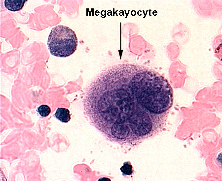
Megakaryocytes are extremely large cells in the bone marrow from which platelets are derived. They are fragile so many are lost in smear preparations, however, when found are easily identified by the large size and multilobed nuclei (Megakaryocyte 1). These large cells are easily identified in bone marrow sections (Megakaryocyte 2).
See if you can identify the developing blood cells in the following fields:
Bone Marrow Smear 1 Answer
Bone Marrow Smear 2 Answer