The female reproductive system includes paired ovaries, which produce female germ cells (oocytes or eggs) and the female hormones estrogen and progesterone. The ovary consists of a cortex, which houses the maturing eggs in follicles, and a medulla, in which blood vessels and nerves enter the structure. Mature eggs are ovulated from the ovary into the oviduct. The egg is received by the funnel shaped infundibulum of the oviduct and travels down the tube through the ampulla, isthmus, and intramural portions of the oviduct to enter the uterus. The uterus is lined by a special mucus membrane, the endometrium, which undergoes cyclic changes in preparation for the implantation of the fertilized egg. The uterus runs continuous with the cervix, which in turn leads into the vagina. The mammary glands are the major accessory glands.
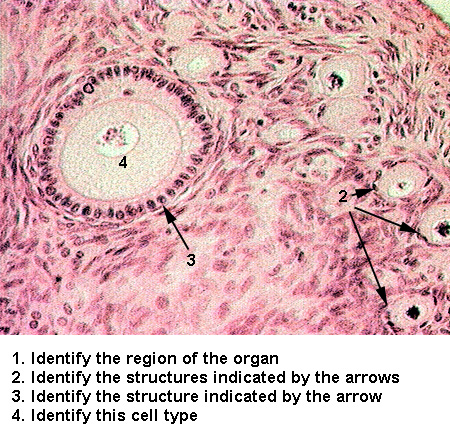
The ovary is covered by cuboidal epithelium. Beneath this epithelium is a band of extracellular matrix materials called the tunica albuginea. The maturing oocytes are surrounded by an epithelial layer, producing a structure called the follicle. Oocytes surrounded by a flattened, single, layer of epithelium are the primordial follicles (Primordial Follicles). As the primordial follicles begin to grow, the surrounding follicular epithelium (or granulosa cells) change morphology. Early primary follicles are surrounded by cuboidal or columnar epithelium (Early Primary Follicle). Late staged primary follicles are surrounded by a stratified follicular epithelium (Late primary follicle). During the primary stage the oocyte becomes surrounded by an acellular layer, the zona pellucida (Zona Pellucida). Secondary follicles form as fluid filled spaces appear in the stratified granulosa cells. These spaces will coalesce into the antral cavity (Secondary Follicle). Surrounding the secondary follicles are two layers of connective tissue, the theca interna, which produces estrogen, contains many cells and capillaries, and the theca externa containing typical dense connective tissue (Thecas). The most mature follicles are Graafianfollicles. The antral cavity has expanded producing a compressed stratified layer of granulosa cells. The oocyte is supported on a thin "stalk" of epithelium, the antrum or cumulus oophorus, and is surrounded by the zona pellucida, and a layer of epithelium, the corona radiata (Graafian Follicle).
After ovulation, the cells of the follicular epithelium and theca interna, under the influence of luteinizing hormone, differentiate into an endocrine gland called the corpus luteum (Corpus Luteum 1). The cells are large, pale staining and the gland is penetrated by delicate connective tissue septa and capillaries (Corpus Luteum 2). This gland produces progesterone which effects the morphology of the uterine endometrium. A degenerated corpus luteum is called the corpusalbicans. It appears as a very pale staining white scar (Corpus Albicans). It may show an accumulation of blood during the early stages of degeneration.
The ovulated egg is captured by the flared oviduct. Via ciliary action and smooth muscle peristaltic contractions, the egg is propelled into the uterus. The bottom of the uterus is constricted to a small opening surrounded by the cerivx, which runs into the vagina.
The oviduct or fallopian tube is the tubule which receives the ovulated egg and transports it to the uterus. It is made of three layers: (1) a mucus membrane, (2) a muscle coat, and (3) a serosa. The mucus membrane is thrown into extensive folds and is composed of a simple columnar epithelium, with ciliated and non-ciliated cells, sitting on a lamina propria of loose connective tissue. It is surrounded by two layers of smooth muscle and a serosa (Oviduct).
The uterus is composed of three layers: (1) a mucus membrane, the endometrium (2) a muscle coat, the myometrium, and (3) a serosa. The endometrium consists of a simple columnar epithelial lining, from which numerous uterine glands extend. The glands penetrate the lamina propria or endometrial stroma. It is this layer which undergoes changes during the female cycle. Compare the following stages:
Estrogenic and Resting phases: - Under influence of estrogen, the endometrial layer regenerates. The layer is fairly thin, the glands straight and narrow, separated by a great deal of dense, cellular endometrial stroma (Resting Endometrium 1 and Resting Endometrium 2).
Progestational or secretory phase: Secretion of progesterone by the corpus luteum prepares the endometrium to accept the fertilized egg. The endometrial lining thickens by the collection of extracellular fluid (edema) producing a lighter staining endometrial stroma. Additionally, the uterine glands begin secretion, dilating and becoming coiled. A basal zone, containing the blind ends of the uterine glands abuts the myometrium and undergoes no change during the female cycle. The stem cells for uterine repair arise from this region (Progestational Endometrium 1 and Progestational Endometrium 2).
The cervix is the lower cylindrical portion of the uterus which projects into the vagina. It consists of a mucus membrane lining deep furrows in a very dense collagenous lamina propria (Cervix 1). The mucus membrane lining the furrow is a simple columnar mucus secreting epithelium forming what appear like tubular glands. These "glands" are called the endocervical glands (Cervix 2). The epithelium of the cervix changes abruptly to stratified squamous non-keratinized epithelium, as it enters the vagina.
The vagina is a musculofibrous tube lined with a mucus membrane of stratified squamous non-keratinized epithelium on a lamina propria. The mucus membrane is surrounded by two layers of smooth muscle, arranged mostly in a longitudinal fashion. The vagina is wrapped by an adventitia (Vagina).
Mammary glands consist of a number of lobes, each of which has a separate duct system. Before pregnancy, the mammary gland lacks secretory units, existing as only a duct system surrounded by a loose connective tissue and adipose tissue (Inactive Mammary Gland). With pregnancy, secretory alveoli appear, consisting of simple columnar cells surrounded by myoepithelium. The proliferation of the secretory units, decreases the amount of connective, condensing it into septa between the lobules (Active Mammary Gland 1). Secretory product often accumulates within the secretory units, dilating them and flattening the epithelium (Active Mammary Gland 2).
See if you can identify the following: