

These pages support the Animal Histology (146:322) course at Rutgers University. Use the following links to other histology sites on the web.
These are the PowerPoint Presentations used in lecture. The information is presented with histological images taken by light microscopy, and augmented with transmission electron microscopy (TEM) and scanning electron microscopy (SEM) images.
These pages contain a number of digitized images for the study of histology. The images have been collected by Bruce Babiarz, PhD, Professor of Cell Biology & Neuroscience and Histology Instructor at Rutgers University. The following tissues and organ systems are available:
The Labs
 Cytology
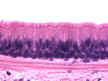 Epithelium
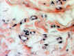 Loose Connective Tissue and Adipose
 Dense Connective Tissue, Cartilage, Bone and Joints
 Blood and Blood Forming Tissues
 Blood Vessels
 Lymphatic System
 Muscle
 Nervous System
 Integument
 Sensory Systems
 Digestive System
 Respiratory System
 Urinary System
 Female Reproductive System
 Male Reproductive System
 Endocrine Systems
Each section is presented with a short descriptive text and links to labeled pictures. At the end of the section the images are presented again, in an unlabeled, lab practical format for review. The thee review sections, covering the material on Lab Practical Exams I, II, and III, each contain 6 practice exams with 10 scopes each.
Visitors since 9/04/01
All images were acquired using a Nikon Diaphot inverted microscope and Image Central Software in the Imaging Facility, Nelson
Biological Labs, Rutgers University. Images were manipulated and labeled using Adobe Photoshop. All images are
copyrighted and not available for general distribution.
Please e-mail any comments to: babiarz@biology.rutgers.edu