The endocrine system is composed of a number of glands, which deposit their secretory products directly into the blood stream. A number of the glands of this system exist as separated individual glands, such as the pituitary, thyroid, parathyroid, adrenal, and pineal body. Others exist as part of other organs or glands, such as the Islet of Langerhans in the pancreas. Although all have different embryological origins, they all share the same basic histological organizations: Groups of cells irregularly organized as cords or clumps, separated by capillaries or blood sinuses.
The pituitary gland is composed of four anatomical parts: (1) the pars distalis or anterior pituitary; (2) pars tuberalis; (3) pars intermedia, and (4) pars nervosa or posterior pituitary (Pituitary).
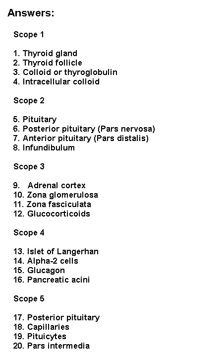
The pars distalis is composed of irregular cords of cells, between which are numerous capillaries. In routine preparations (H&E) some of the cells stain (chromophils = actively secreting) and some do not (chromophobes) (Anterior Pituitary 1). In sections stained with trichromes, chromophils can be further divided into acidophilic and basophilic cell types (Anterior Pituitary 2). The pars tuberalis projects off the pars distalis and is composed of cuboidal cells and a large number of blood vessels. It represents the entrance of the hypophysioportal blood system into the anterior pituitary. The pars distalis is separated from the pars nervosa by a cleft or row of follicles. Immediately adjacent to this division, associated with the pars nervosa, is the pars intermedia. It is composed of pale staining cells, arranged in follicles, or as a few rows of basophilic cells and associated capillaries (Pars Intermedia). The pars nervosa or posterior pituitary is composed of the axonal projections of the hypothalamohypophyseal tract. In a good preparation, the terminals of the axons can be seen abutting capillaries (Posterior Pituitary). In this region, secretory product accumulates around the end bulb, forming the palisades zone. Other larger bodies, the Herring bodies, which also represent the accumulation of secretory products in axon terminal bulbs, can be seen. The nuclei observed in the pars nervosa, are those of the supporting cell type, the pituicyte.
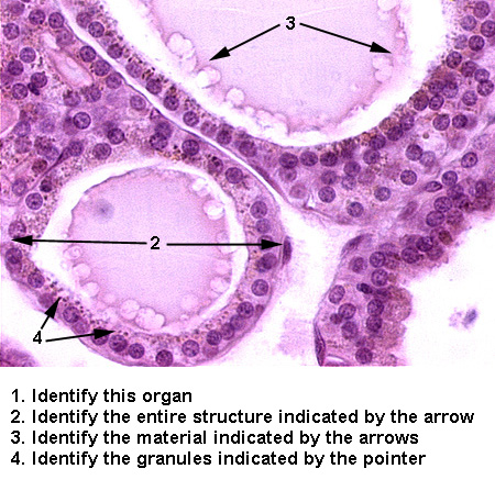
The cells of the thyroid gland are organized into structural and functional units called follicles (Thyroid 1). The follicular cells are arranged as a simple cuboidal epithelium with a basement membrane, surrounding a central mass of stored precursor, the colloid ot thyroglobulin. Granules within the cells represent intracellular collaoid (Thyroid 2). A second cell type, the parafollicular or C cells, sits on the outside of the follicle (C-cells). The follicular cells produce thyroxine; the parafollicular cells produce calcitonin.
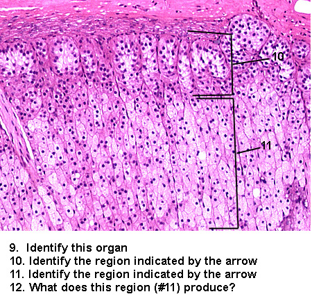
The parathyroid gland consists of two paired glands lying on each side of the thyroid. The cells are arranged in irregular cords, supported by reticular fibers, and abut numerous capillaries (Parathyroid 1). Two types of cells are present: (1) chief or principle cells and (2) oxyphil cells. The chief or principle cells are the major cell type and are small cells with spherical nuclei, and have pale staining, finely granular cytoplasm. The oxyphil cells are found in clumps, usually at the periphery of the gland. They are much larger than the chief cells, are very pale staining, and have distinct cell boundaries (Parathyroid 2).
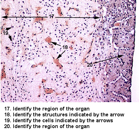
The adrenal or suprarenal glands lie in contact with the upper surface of the kidney. They consist of two regions, the cortex and medulla (Adrenal Gland). The cortex is composed of three layers: (1) Zona glomerulosa, located beneath the connective tissue capsule, consists of irregular clusters of columnar cells (Adrenal cortex 1). (2) Zona fasciculata, the thickest layer, located beneath the zona glomerulosa, consists of straight cords of cells perpendicular to the surface. These cells, called spongiocytes, have very high cholesterol content and appear light staining (Adrenal cortex 2). (3) Zona reticularis, the inner most layer, consists of thin anastomosing cords of cells with increased acidophilic staining (Adrenal cortex 3). The medulla occupies the center of the gland and consists of large ovoid basophilic cells arranged in clumps or irregular cords around an extensive capillary system. This region also contains numerous veins (Adrenal medulla).
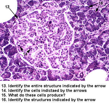
The Islets of Langerhans are found scattered between the exocrine acini of the pancreas (Islet 1). The cells are arranged in clumps between capillaries. Four different cell types are present, however, they rarely can be distinguished without special stains. The majority of the cells are the insulin secreting beta cells. The alpha cells, which produce glucagon, can be identified by their darker staining nuclei and more eosinophilic cytoplasm (Islet 2).
The pineal body develops from and is connected to the third ventricle of the brain. It is covered by the pia mater, which sends septa into the gland, dividing it into incomplete lobules (Pineal 1). Associated with rich capillary supply, are two cell types: (1) pinealocytes, which are the major cell type, are found in clumps and highly branched and (2) neuroglial supporting cells, distinguished by their flattened nuclei. In the adult, there is an accumulation of calcified material, known as brain sand (Pineal 2).
See if you can identify the following: