Epithelia are grouped into two general classes: (1) Epithelial Membranes - covering or lining of the body (skin or stomach lining) and (2) Glandular Epithelium - for increased secretion epithelial cells grow down into the connective tissue and form glands.
Epithelial membranes provide many diverse functions in the body including protection from wear and tear, drying, or excess moisture; secretion of many different products; and absorption of nutrients and other molecules. Therefore epithelium comes in a variety of forms adapted for a particular function. Find three basic types of epithelial membranes:
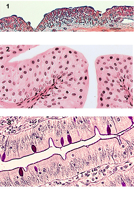
Simple squamous epithelium - cells are flat ("scale-like") in single layer supported by an underlying basement membrane (BM) composed of connective tissue molecules. It is found in 2 basic areas: (1) mesothelium which lines all body cavities & covers all organs (Squamous 1) and Squamous 2) and (2) endothelium which makes up the walls of capillaries and the inner lining of blood vessels (Squamous 3).
Simple cuboidal epithelium - sectioned at right angle (cross-section) the cells appear cube shaped (Cuboidal 1). This cell type is found primarily in duct systems (Cuboidal 2) or as the collecting tubules in kidney (Cuboidal 3).
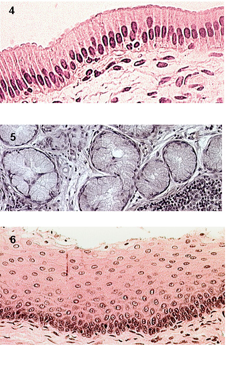
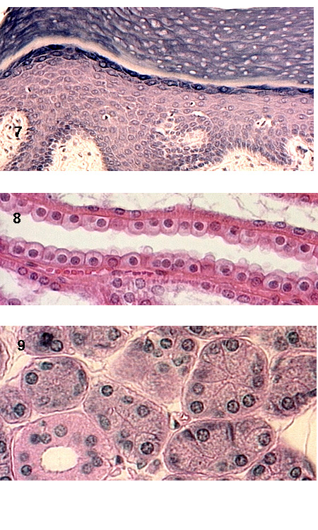
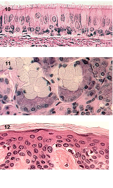
Simple columnar epithelium - cells taller than wide w/ many subtypes. It forms most of the large ducts in the body (Columnar duct) and the lining of absorptive cells in the gut (Columnar Absorptive 1). At higher magnification, absorptive cells possess a "brush border" or extensive microvilli at their surface (Columnar Absorptive 2). In some case, such as the stomach lining, all of the cells have become mucus secreting cells (Columnar Mucus 1). Mucus secreting cells have a characteristic morphology described as "frothy" appearing cytoplasm (Columnar Mucus 2). Within parts of the intestine the simple columnar membrane provides two functions, secretion of mucus and absorption of nutrients. Thus the membrane consists of two different cell type, absorptive cells with microvilli and goblet cells, which secrete mucus (Columnar w/ Goblets 1). Using the PAS staining technique these goblet cells stain a bright fuschia, and their secreted product visible on the surface of the membrane (Columnar with Goblets 2).
Pseudostratified epithelium is found primarily in the respiratory system and is often referred to as "Respiratory Epithelium". Its organization is described as a membrane in which some cells contact BM and don't reach surface, and some do (Pseudostratified with Cilia 1). In most cases this membrane is composed of ciliated cells with intermixed goblet cells (Pseudostratified with Cilia 2).
Stratified squamous non-keratinizing epithelium - This epithelium is found covering wet surfaces which receive significant "wear and tear" - e.g. inside mouth, esophagus, anal canal. It is NOT composed of successive layers of squamous cells, but consists of basal columnar, polymorphic, and surface squamous layers (Stratified Squamous non-keratinized 1).
Transitional epithelium - This epithelium lines the expandable tubes and organs of the body - e.g. bladder. When the bladder is full and the membrane is stretched, it looks like stratified squamous non-keratinized epithelium. When relaxed, the surface cells obtain a rounded morphology (Transitional Relaxed 1).
Stratified squamous keratinizing epithelium - This epithelium has the same morphology as stratified squamous non-keratinized, but with the outer layer undergoing a metamorphosis to tough non-living layer of keratin. It will form the outer layer of the skin (epidermis) and be either thin (Stratified Squamous Keratinized 1) or thick (Stratified Squamous Keratinized 2). In these pictures, two distinctive features are observed, a layer of diamond shaped cells possessing keratohyaline granules and the surface keratin layer.
There are two main types of glands in the body: exocrine - which convey secretions via ducts and endocrine - which secrete directly into body (capillaries). Endocrine glands will be covered later. This section will detail the types of exocrine glands. These glands contain two epithelial components, secretory units and ductal epithelium. They are classified by many different factors:
Serous Glands - look like a pie cut in sections, with cells having round nuclei and basophilic cytoplasm (Serous Gland 1 and Serous Gland 2).
Mucus Glands - have flat nuclei at the base of each cell with frothy, pale cytoplasm (Mucus Gland 1).
Mixed Glands - consist of mucus gland units as above with associated a crescent of serous cells, a "serous demilune" (Mixed Gland 1 and Mixed Gland 2). Both mucus and serous secretions share a common duct system in these glands.
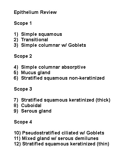
See if you can identify the epithelial type in each of the following:
Scope 1 Scope 2 Scope 3 Scope 4 Answers