The urinary system eliminates waste products from the body and maintains fluid/salt balance. The system consists of paired kidneys with ureters, a urinary bladder, and urethra.
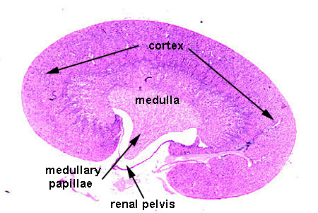
The kidney is covered by a thin connective tissue capsule and consists of an outer cortex and medullary pyramid or papillae (Kidney). Within these two regions are found the components of the structural and functional unit of the kidney, the nephron. The nephron is composed of: (1) the glomerulus, a tuft of capillaries, which produces the glomerular filtrate, housed in the renal corpuscle; followed by a series of tubules, specialized for excretion and reabsorption, including (2) the proximal convoluted tubule, (3) the descending and ascending loop of Henle, and (4) the distal convoluted tubule. Each nephron drains into a collecting tubule, which serves as a duct system to conduct the urine out of the Kidney. The glomeruli and the proximal and distal convoluted tubules are found in the cortex. The descending loop of Henle leaves the cortex and enters the medulla, returning to the cortex as the ascending loop. Therefore, the medulla consists of portions of the loops of Henle and the collecting tubules.
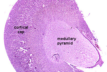
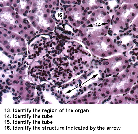
The outer most region of the kidney, which lies just below the convex surface of the organ, is the cortex, where three components of the nephron can be found. The renal corpuscles (or Bowman's capsules) containing glomeruli are surrounded by a labyrinth of proximal and distal convoluted tubules (Cortex). The collecting tubules also penetrate the cortex, to connect with the distal convoluted tubules. These extensions are called medullary rays and represent the cores of the kidney lobules (Medullary Rays).
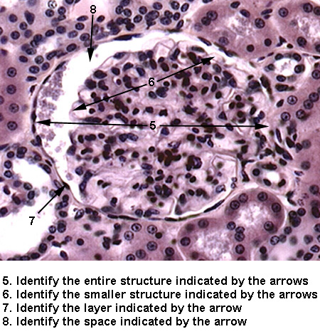
The glomeruli consist of a tuft of capillaries housed in an epithelial lined Bowman's capsule (Renal Corpuscle). The parietal epithelium of Bowman's Capsule is a layer of simple squamous epithelium lining the outer border of the corpuscle. The visceral epithelium of Bowman's capsule (or podocytes) surrounds the capillary endothelial cells, with mesangial cells filling in the spaces between closely apposed capillaries. The afferent arteriole enters the corpuscle and the efferent arteriole leaves the corpuscle, both at a region called the vascular pole.
The glomerular filtrate leaves the corpuscle via the proximal convoluted tubule. The proximal convoluted tubule travels a tortuous course, therefore will appear as tubes cut in various orientations (i.e. cross-sectional or oblique). They are the most prominent tubule seen in the cortex. The cells stain highly acidophilic and possess a brush border (Proximal Tubules). The distal convoluted tubules differ from the proximal tubules in that: (1) the total diameter of the tubule is smaller, (2) but, the cells are lower producing a larger lumen, (3) the cells are less acidophilic, and (4) the cells do not have a brush border (Distal Tubules). The distal tubules also travel a tortuous course, producing different cut orientations. Before joining the collecting tubules, the distal convoluted tubules abut the renal corpuscle at the vascular pole. At this site, the distal tubule wall has an increased number of nuclei, producing a structure called the macula densa (Macula Densa).
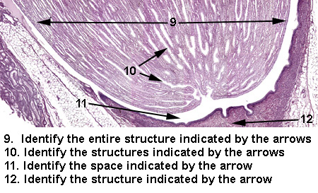
The medullary portion of the kidney is organized as a single medullary pyramid (unilobar kidney) or multiple pyramids (multilobar kidney). Each pyramid of medullary tissue and its associated "cap" of cortical tissue is defined as a kidney lobe (Kidney Lobe). The pyramids appear striated, due to the parallel alignment of the loops of Henle and collecting tubules (Medulla 1). Histologically, the loop of Henle appear acidophilic as the descending/ascending loop (Medulla 2) and becomes a thin squamous lined tube near the tip of the papillae (Medulla 3). Collecting tubules are not considered part of the nephron; they are the duct system of the kidney. The bulk of the medullary pyramid is composed of collecting tubules. The collecting tubules are lined with simple cuboidal epithelium. They meet at the apex or papillae of the medullary pyramid, merging together to form large ducts, the ducts of Bellini which empty into the renal pelvis (Papillae).
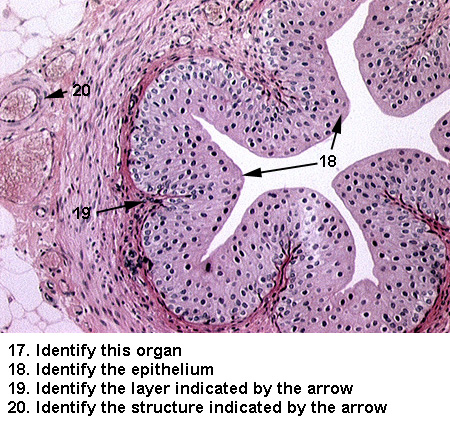
The urine in the collecting tubules is collected in the renal pelvis and exits the kidney in the ureter. The ureter travels to the bladder, where the urine can be stored. The bladder is drained by the urethra which leads to the external orifice.
The ureter is composed of a folded mucus membrane, a muscle coat, and a fibroelastic adventitia (Ureter 1). The mucus membrane consists of two layers: (1) transitional epithelium and (2) lamina propria (Ureter 2). The muscle coat consists of two layers of smooth muscle.
The urinary bladder is lined with transitional epithelium underlined by a collagenous lamina propria. A submucosa of elastic fibers and a muscular layer of three coats of smooth muscle permit expansion of the structure (Bladder).
In the male, the urethra runs within the prostate gland and penis, and will be studied in the male reproductive section. In the female, it is a separate tube consisting of a mucus membrane (epithelium and lamina propria), submucosa and muscular coat of two layers of smooth muscle. The epithelium varies: transitional by the bladder, changing to stratified squamous non-keratinizing (Female Urethra), and finally stratified squamous at the opening.
See if you can identify the following: