The lymphatic system is responsible for the production and storage of the agranular white blood cells or lymphocytes. There are two types of lymphocytes, the T-cells, which are involved in cell mediated defense mechanisms of the body, and B-cells, which are involved in the humoral response. Lymphatic tissue is found in four different forms in the body:
These tissues are composed of two principle components (1) free cells (lymphocytes), and (2) a supporting frame work of extracellular fibers (reticular) and the fibroblasts which produce them.
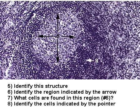
This consists primarily of lymphatic nodules, which are sites of lymphocyte (B-cells)
proliferation/activation. The basic structure of a lymphatic nodule is described as a germinal
center, where B-lymphocytes have activated and increased in size, surrounded by small,
inactivated and proliferating B-cells (Lymphatic Nodule). These lymphatic nodules cab form
anywhere in the body when B-cells are activated by their specific antigen. Some have permanent
localizations, such as Peyer's Patches in the submucosa of the ileum (Peyer's Patches). In
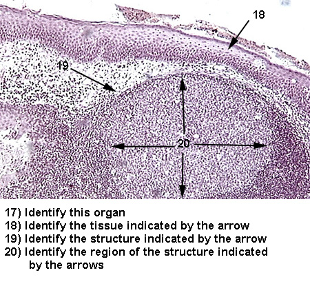
defending the nasal and oral cavities of the body, many of these lymphatic nodules become
grouped together forming tonsils. The structure of tonsils basically include a group of lymphatic
nodules covered by an epithelium and basement membrane. There are three large paired tonsils
in the body: Palatine tonsils and Lingual tonsils with a stratified squamous non-keratinized
epithelial covering (Palatine/Lingual Tonsil) and Pharyngeal tonsils covered with a
pseudostratified epithelium (Pharyngeal Tonsil).
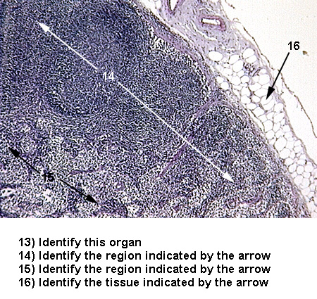
Lymph nodes are found throughout the body associated with the lymphatic vessels, and function
to filter the lymph. lymph nodes are surrounded by a connective tissue capsule and are usually
found embedded in fat. Their structure includes a number of lymphatic nodules in the outer
cortex region (Lymph Node 1) and a central medulla containing irregular cords of circulating B-
and T-cells supported by reticular fibers (Lymph Node 2).
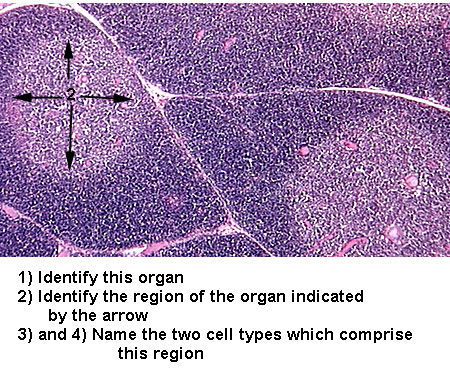
This organ has a thick connective tissue capsule and thick connective tissue septa. The bulk
(pulp) of the organ is composed of two types: white pulp and red pulp. The white pulp is
composed of lymphatic nodules and the red pulp consists of irregular chords of cells and blood
sinuses lined with endothelial cells (Spleen 1 and
Spleen 2). The red pulp is the region of the
spleen which functions to filter the blood, removing worn out RBCs with the help of
macrophages.
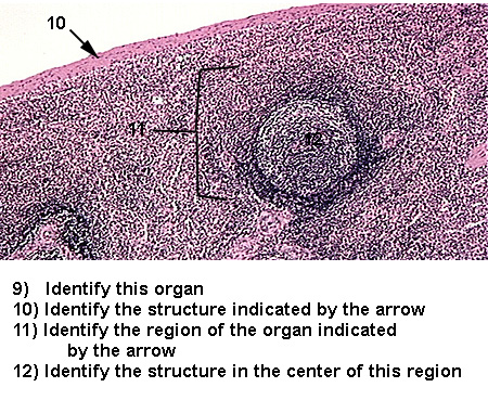
The thymus is the site of T-cell production. The organ is bilobed and each lobe is divided into a number of lobules by connective tissue septa. Each lobule has a dense concentration of immature T-lymphocytes at the periphery, the cortex, and a central part, the medulla, where circulating mature T-lymphocytes enter the organ (Thymus 1). In both regions the T-cells are supported by epithelioreticular cells, identified by their larger, lighter staining nuclei (Thymus 2). Within the medullary regions one can identify thymic corpuscles (Hassall's corpuscles) which appear as spherical or ovoid bodies composed of concentrically arranged epithelial cells (Thymus 3). No lymphatic nodules are seen in either area.
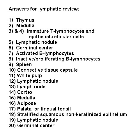
See if you can identify the following lymphatic tissues:
Scope 1 Scope 2 Scope 3 Scope 4 Scope 5 Answers