The respiratory system consists of two functional parts: (1) a conducting system to bring the air in, including the nose, pharynx, larynx, trachea, bronchi and bronchioles; and (2) the respiratory portion, the site of gas exchange, including the respiratory bronchioles, alveolar ducts, alveolar saccules, and alveoli. The conducting system exists both outside (nose to bronchi) and within (bronchi and bronchioles) the lungs. To prevent collapse upon inspiration the walls of the conducting tubes are supported by cartilage. To strain, warm (or cool), and humidify the air the tubes are lined by a mucus membrane.
Nose - Nasal Cavities
The nose consists of two nasal cavity separated by a cartilaginous nasal septum which is covered with a mucus membrane (Nasal Septum). Air enters through the nostrils. which are openings lined by an thin skin with coarse hairs and sebaceous glands. Inside the cavities, the epithelium changes to stratified squamous non-keratinized within the vestibules and to pseudostratified ciliated columnar type with goblet cells in the inner respiratory region. The epithelium sits on a lamina propria, containing numerous serous and mucus glands (Nasal Mucus Membrane). The nasal cavities are also lined in part by the olfactory epithelium, described in detail in the sensory laboratory (Olfactory Epithelium). Projecting from the wall of the nasal cavity a boney shelves called the nasal conchae (Nasal Conchae 1). The conchae are covered by respiratory epithelium (pseudostratified ciliated columnar with goblet cells) with a lamina propria containing numerous, collapsed veins (Nasal Conchae 2).
Air moves through the nasal cavities and into the pharynx. The pharynx forms the back of the oral cavity and possesses a histology similar to that of the cheek. It consists of a mucus membrane composed of stratified squamous non-keratinized epithelium resting on a lamina propria containing numerous mucus glands (Cheek).
The epiglottis is a flap-like projection, covered by a mucus membrane, which sits on top of the larynx (Epiglottis 1). The epithelium is stratified squamous non-keratinized except for a smaller circular patch of respiratory epithelium which sits in the laryngeal opening. The epiglottis has a core of elastic cartilage and numerous mixed glands are found in the lamina propria on the laryngeal side (Epiglottis 2).
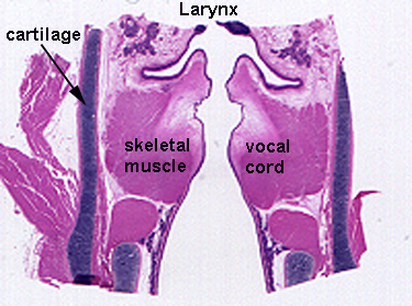
The larynx connects the pharynx and trachea. The tube contains several hyaline cartilages, bound together by connective tissue, to prevent collapse and contains the vocal cords which function in phonation (Larynx 1). It is lined with respiratory epithelium (pseudostratified ciliated columnar) except on the vocal chords which are covered by stratified squamous non-keratinized epithelium (Larynx 2). The vocal chords are folds in the mucus membrane, covered by stratified squamous non-keratinized epithelium under laid with a layer of fibroelastic connective tissue, with a core of skeletal muscle (Vocal Cords).
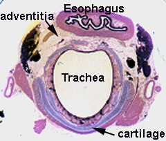
The trachea connects the larynx and bronchi. It is lined with a mucus membrane with respiratory epithelium and a lamina propria, and a submucosa containing horseshoe shape hyaline cartilages and numerous mucus and mixed glands (Trachea 1). The gap between the ends of the cartilages is bridged by fibroelastic connective tissue and smooth muscle (Trachea 2). To increase expandability, the respiratory epithelium is underlaid with a lamina propria containing a distinct elastic lamina (Trachea 3). The trachea is surrounded by an adventitia which is a connective tissue layer shared with the esophagus (Trachea 4).
The trachea ends by dividing into two branches, the 10 or extrapulmonary Bronchi. These tubes have the same histology as the trachea. They enter the lungs at the hilus and branch to each lobe as 2o or Lobar Bronchi. These will branch and enter the lungs within septa as 30 or Segmental Bronchi. Their composition is the same as the primary bronchi except the walls contain irregular shaped cartilaginous plates and a complete layer of smooth muscle around the tube (Bronchi 1). The respiratory epithelial lining often appears folded due to the contraction of the smooth muscle, and mucus glands are found between the muscle and cartilage (Bronchi 2).
The bronchioles branch off segmental bronchi and penetrate into the lung lobules. The preterminal bronchioles are the first branches off bronchi and continue branching into smaller tubes called terminal bronchioles . The preterminal bronchioles consist of a folded layer of simple columnar ciliated epithelium (Preterminal Bronchioles 1), whereas in terminal bronchioles the epithelial layers is flattened (Terminal Bronchioles 1). These tubes contain no cartilage, goblet cells, or mucus glands. The epithelium contains non-ciliated Clara Cells which secrete a surfactant, supported by a lamina propria, and smooth muscle layer (Preterminal Bronchioles 2). The walls of the bronchioles become discontinuous, with the appearance of outpocketings in walls which lead to areas of gas exchange. These tubes are considered to be the respiratory bronchioles (Terminal Bronchioles 1). These tubes have free terminations which open into straight spaces in the respiratory portion called alveolar ducts (Respiratory Bronchioles 2).
The "structures" of the respiratory portion of the lung have no walls of their own, but are spaces - the alveolar ducts, saccules, and alveoli - in a huge elastic sponge-like capillary bed (Respiratory Portion). The alveolar ducts are straight spaces continuous with the free terminations or the respiratory bronchioles (Alveolar Ducts). The alveolar saccules are rotunda-like spaces communicating with the ducts and the alveoli are spur-like partitions around the periphery of the saccules (Saccules). In the respiratory portion of the lung, the cellular composition is referred to as the interalveolar wall. The walls are composed of capillary endothelial cells and a lining of epithelium. Two epithelial cell types are present; (1) pneumocyte type I, a very thin squamous cell type, which functions in gas exchange, and (2) pneumocyte Type II, a rounded cell with cytoplasmic granules, which functions to produce a pulmonary surfactant (Interalveolar Wall 1). The details of the interalveolar wall are hard to discern in regular histology sections (7µm), but in thin sections (1µm) pneumocyte types are easily identified (Interalveolar Wall 2). At the surface of the wall, or in the free space, alveolar phagocytes or dust cells can be seen (Interalveolar Wall 3).
Review